Many patients in the early stages of aortic stenosis misinterpret their symptoms as a normal part of ageing. As a result, breathlessness, fatigue, reduced stamina, or slower recovery after activity are often overlooked, delaying recognition of a progressive cardiac condition that requires medical attention. This rationalisation is reinforced by gradual symptom onset and the absence of early pain, often leading individuals to adapt their lifestyles rather than seek assessment. As a result, medical review is frequently delayed, allowing the disease to advance silently until symptoms become more disruptive, persistent, and clinically significant. By the time a diagnosis is made, intervention is often more urgent and complex, carrying higher health risks, fewer treatment options, and less flexibility in selecting the most appropriate therapeutic approach.
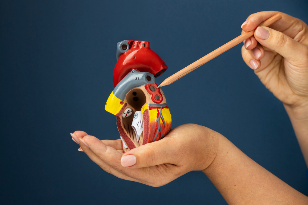
The biology: the narrowing of the aortic gateway
The aortic valve acts as the heart’s primary gateway, allowing oxygenated blood to pass from the left ventricle into the aorta and onwards to the rest of the body. In a healthy heart, this valve consists of three thin, flexible leaflets that open fully with each heartbeat and close securely to prevent backflow. With aortic stenosis, these leaflets gradually become thickened, stiffened, and calcified, losing their ability to open freely and efficiently during systole, thereby disrupting normal blood flow dynamics.
Age-related calcium deposition is the most common cause, although congenital valve abnormalities and previous inflammatory conditions can also contribute. As calcification progresses, the effective valve opening becomes smaller, increasing resistance to blood flow leaving the heart. This narrowing forces the left ventricle to work harder to maintain adequate circulation, placing sustained strain on the cardiac muscle over time and accelerating structural changes that compromise performance.
As the valve narrows further, the left ventricle must generate significantly higher pressure to eject blood through the restricted opening. This chronic pressure overload causes the muscular wall of the ventricle to thicken, a process known as left ventricular hypertrophy. While initially compensatory, this thickening reduces ventricular compliance, increases oxygen demand, and eventually compromises filling and pumping efficiency, setting the stage for symptomatic disease, heart failure, and poorer long-term prognosis without timely intervention.
Strategy 1: identifying the “SAD” triad of symptoms
Clinical recognition of aortic stenosis often relies on awareness of the classic symptom cluster known as the “SAD” triad (syncope, angina, and dyspnoea). These symptoms typically emerge as the condition progresses from moderate to severe, indicating that the obstruction has reached a level where the heart can no longer compensate effectively during physical or physiological stress, particularly during exertion or periods of increased demand.
Syncope refers to episodes of fainting or near-fainting, most commonly occurring during exertion or sudden changes in posture. Physical activity increases the body’s demand for blood flow, yet the narrowed valve restricts the heart’s ability to respond appropriately. This mismatch can result in a temporary reduction in blood supply to the brain, causing dizziness, light-headedness, or loss of consciousness, which should always prompt urgent clinical evaluation and specialist assessment.
Angina, or chest pain, occurs when the thickened heart muscle requires more oxygen than the coronary circulation can supply. Even in patients without coronary artery disease, the increased workload and reduced perfusion reserve of the hypertrophied ventricle can lead to chest discomfort. This pain is often exertional, pressure-like in nature, and may resolve with rest as myocardial demand decreases, potentially masking its cardiac origin.
Dyspnoea, defined as shortness of breath on exertion, reflects rising pressures within the heart and lungs. Impaired ventricular filling leads to increased pressure in the left atrium and pulmonary circulation, causing fluid congestion. Over time, activities such as walking uphill, climbing stairs, or carrying groceries become increasingly difficult, reducing independence, exercise tolerance, and overall functional capacity in daily life.
Strategy 2: the Significance of the heart murmur
A heart murmur is frequently the earliest objective sign of aortic stenosis and is often detected during routine clinical examination. General practitioners play a pivotal role in early identification, as careful auscultation can prompt timely referral for further investigation before symptoms become advanced or complications such as heart failure, arrhythmias, or sudden deterioration develop. The characteristic murmur associated with aortic stenosis is described as an ejection systolic murmur. It is generated by turbulent blood flow as blood is forced through the narrowed valve during ventricular contraction. Typically heard best at the upper right sternal edge, the sound may radiate to the carotid arteries in the neck, reflecting altered haemodynamics within the aorta and reduced valve opening.
While the loudness of the murmur does not always correlate with disease severity, its presence is clinically significant, particularly when accompanied by exertional symptoms or risk factors such as advanced age. The murmur serves as an audible marker of altered valve function and highlights the importance of regular cardiovascular examinations as part of routine preventive healthcare and ongoing patient monitoring.
Strategy 3: moving from diagnosis to treatment
Once aortic stenosis is suspected, echocardiography is the definitive diagnostic investigation. This non-invasive ultrasound assessment provides detailed visualisation of valve anatomy, leaflet mobility, and blood flow patterns. By measuring pressure gradients across the valve and calculating the valve area, clinicians can accurately classify disease severity and determine the appropriate timing of intervention, ensuring treatment is neither delayed nor premature.
Echocardiography also evaluates left ventricular size, wall thickness, and systolic function, offering insight into the heart’s adaptive response to chronic pressure overload. These findings help guide clinical decision-making, particularly in determining whether symptoms are attributable to valve disease and whether intervention is likely to improve survival, functional capacity, and long-term quality of life.
Historically, surgical aortic valve replacement through open-chest surgery was the standard treatment for severe symptomatic disease. Although effective, it carries a higher procedural risk and prolonged recovery for some patients. The advent of transcatheter aortic valve implantation, or TAVI, has revolutionised management by allowing valve replacement via a catheter-based approach, avoiding sternotomy and enabling faster recovery with fewer complications and reduced hospital stays.
Quality of life post-treatment
Following successful valve replacement, most patients experience a significant and sustained improvement in quality of life. Relief from breathlessness and chest discomfort often occurs within weeks, allowing individuals to resume activities that had previously been limited or avoided due to symptoms, fear, or physical exhaustion and declining confidence, while regaining a greater sense of control over their daily routines and physical capabilities. Physical activities such as walking, gardening, and light exercise typically become easier as cardiac output improves and pulmonary congestion resolves. Many patients also report better functional outcomes, sleep quality, improved mood, and increased confidence in their physical abilities, contributing to overall well-being, independence, and renewed social engagement.
Shortness of breath should not be dismissed as a normal part of getting older. It can be an early warning sign of aortic valve disease, where delayed diagnosis may lead to serious complications. Ongoing follow-up is essential to monitor valve function, manage comorbid conditions, and optimise long-term outcomes. Cardiac rehabilitation and clear lifestyle guidance play an important role in supporting recovery and maintaining functional capacity. Timely treatment of aortic stenosis not only improves symptoms but also significantly enhances survival, highlighting the need for early assessment, accurate diagnosis, and patient-centred intervention supported by continuous clinical oversight. Book a valve assessment today to identify potential valve disease early, before symptoms progress.