The UK's National Lead for Structural Heart Intervention - Now Practicing in London

Heart care delivered by a world-renowned UK cardiologist.

Trusted by patients worldwide, we deliver personalised cardiac care with unmatched expertise. Combining cutting-edge innovation and compassionate support, your heart health is always in the best hands.

Symptoms

Understand the warning signs your heart may be giving you. Explore common symptoms like chest pain, breathlessness, and palpitations — and learn when to seek expert care.

Conditions

Learn more about common and complex heart conditions, from arrhythmias to coronary artery disease. With world-leading expertise, we guide you to the right diagnosis and advanced treatment options.

Treatments

Discover a comprehensive range of advanced cardiac treatments, from state-of-the-art diagnostics to minimally invasive procedures. Each approach is personalised to protect, treat, and strengthen your heart.

About Dr Suneil Aggarwal

A man with short dark hair, wearing blue-rimmed glasses, a navy blazer, and a white collared shirt, poses against a plain white background.

Dr Suneil Aggarwal is an Interventional Cardiologist specialising in valve and congenital heart disease interventions (structural intervention), as well as general cardiology. He qualified with multiple distinctions and prizes from University College London in 2002 and completed his postgraduate training in Oxford, India (Puttaparthi) and London.

He completed an MD research degree from University College London and a 2 year fellowship in TAVI and congenital interventions at The Heart Hospital and Barts Heart Centre in 2015. He has been practicing as Lead for Structural Intervention in Liverpool for the last 9 years, where he developed the service into one of the largest in the UK. He has now relocated to Barts Heart Centre to practice at what is one of the largest cardiac centres in Europe. He has particular expertise in TAVI, mitral interventions, PFO/ASD closure and other complex congenital and valve interventions. He is actively involved in research and training of the next generation of interventional cardiologists, and is regularly invited to speak around the world. As well as this, he regularly participates in voluntary work abroad. His reputation amongst his peers is reflected in being thrice elected to the Council of the British Cardiovascular Intervention Society, the national society for interventional cardiologists. He is now the National Lead for Structural Intervention, which includes PFO closure, TAVI and other valve interventions and congenital heart disease interventions.

Symptoms

Our Client Reviews

Conditions

Diagnostic Tests

State-of-the-art diagnostic testing to accurately assess your heart health and guide treatment decisions.

Treatments

Evidence-based cardiac treatments focused on long-term heart health.

Our Blogs

Understanding Syncope (Fainting)

Syncope (pronounced sin-co-pee) is the medical word for fainting. It describes a brief loss of consciousness caused by a temporary drop in blood flow to the brain. Most episodes last only a few seconds to a couple of minutes, and people usually recover quickly. Although fainting can be frightening, it is often harmless — but occasionally it can be a sign of something more serious.

What Causes Syncope?

There are several main causes of fainting. The key question doctors ask is: Why did blood flow to the brain suddenly drop?

1. Reflex (Vasovagal) Syncope – The Most Common Type

This is the type many people experience at some point in their lives. It can be triggered by:

  • Emotional distress
  • Seeing blood
  • Pain
  • Standing for a long time
  • Dehydration

In these situations, the heart rate slows and blood pressure drops suddenly. You may feel warning symptoms such as dizziness, nausea, sweating, blurred vision, or “tunnel vision” before passing out.

2. Postural (Orthostatic) Hypotension

This happens when blood pressure falls after standing up too quickly. It is more common in older adults, people who are dehydrated, or those taking certain medications (such as blood pressure tablets or diuretics).

3. Cardiac Causes

Fainting related to the heart is less common but more serious. It can occur due to:

  • Abnormal heart rhythms (too fast or too slow)
  • Heart valve problems (such as aortic stenosis)
  • Structural heart disease

Cardiac syncope may happen suddenly without warning, especially during exertion. This type needs urgent medical evaluation.

How Is Syncope Investigated?

If you faint, your doctor will start by asking detailed questions:

  • What were you doing at the time?
  • Were there warning symptoms?
  • How long were you unconscious?
  • Was there any shaking or injury?

A careful history often gives the diagnosis.

Common tests include:

  • Electrocardiogram (ECG) – records the heart’s electrical activity.
  • Blood pressure measurements, including lying and standing readings.
  • Blood tests – to check for anaemia or other issues.
  • Heart monitoring (Holter monitor or event recorder) if an abnormal rhythm is suspected.
  • Echocardiogram (heart ultrasound) if structural heart disease is possible.
  • Occasionally, a tilt-table test, which recreates the faint in a controlled setting to confirm reflex syncope.

Most people do not need extensive testing if the cause is clearly vasovagal and there are no high-risk features.

How Is Syncope Treated?

Treatment depends entirely on the cause.

For Reflex (Vasovagal) Syncope:

  • Stay well hydrated.
  • Avoid triggers where possible.
  • Learn to recognise warning symptoms.
  • If you feel faint, lie down and raise your legs or sit and put your head between your knees.
  • Some people benefit from physical counter-pressure manoeuvres (such as crossing legs and tensing muscles).

Medication is rarely needed.

For Orthostatic Hypotension:

  • Drink more fluids.
  • Stand up slowly.
  • Review medications with your doctor.
  • Compression stockings may help in some cases.

For Cardiac Syncope:

Treatment may include:

  • Pacemaker insertion (for slow heart rhythms)
  • Medication
  • Catheter ablation (for certain fast rhythms)
  • Valve intervention or other heart procedures

When Should You Seek Urgent Help?

Seek medical attention promptly if fainting:

  • Happens during exercise
  • Occurs without warning
  • Is associated with chest pain, increasing shortness of breath or palpitations
  • Happens in someone with known heart disease
  • Results in significant injury

Summary

Most fainting episodes are benign and manageable, especially in young healthy individuals. However, it is always worth discussing syncope with a healthcare professional to rule out more serious causes. With the right assessment, most people can be reassured — and helped to prevent future episodes. If a serious cause is found (usually cardiac), then of course this can usually be treated.

A patient’s guide to cardiac MRI: what to expect

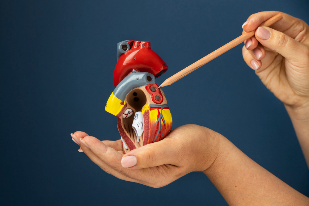
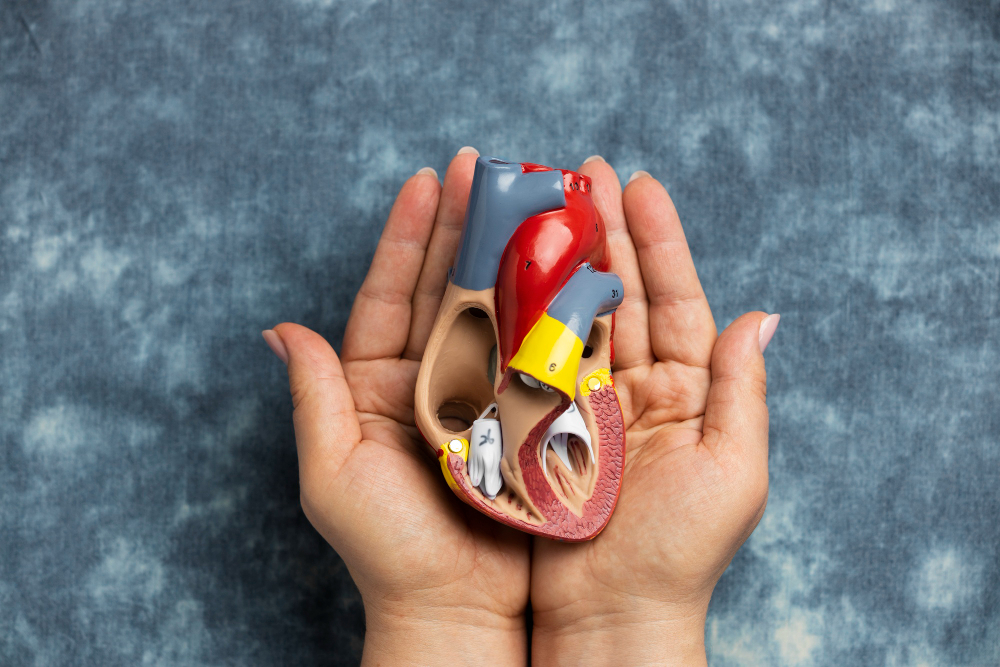
If an ultrasound is a sketch of your heart, a cardiac MRI is a high-definition 3D model. It offers an advanced, non-invasive way to visualise the heart’s structure, movement, and tissue health with exceptional clarity. Using powerful magnetic fields and sophisticated imaging techniques, it enables clinicians to examine the heart from multiple angles and assess the condition of the heart muscle at a microscopic level. This depth of information is particularly important when symptoms are unexplained, conditions are complex, or previous investigations have not provided definitive answers. Cardiac MRI has become an essential tool in modern cardiology, supporting accurate diagnosis and personalised treatment planning. This guide explains what the scan involves, why it is recommended, and how patients can prepare with confidence, ensuring a clear understanding of the process from start to finish.

The biology: magnetic resonance vs ultrasound

Cardiac MRI and ultrasound both assess heart health, but they operate on fundamentally different principles that influence the type and depth of information they provide. Ultrasound uses high-frequency sound waves to create real-time moving images of the heart, making it particularly effective for evaluating valve function, chamber size, and blood flow dynamics during each heartbeat. However, its ability to visualise deeper structures is limited by bone, lung tissue, body shape, and variability in image quality between patients. Magnetic resonance imaging, by contrast, uses a strong magnetic field combined with radiofrequency signals to generate highly detailed images of the heart in multiple planes, offering consistent image quality and comprehensive anatomical assessment.

The key advantage of cardiac MRI is tissue characterisation, a capability that extends beyond structural imaging. It can differentiate healthy heart muscle from scarred, inflamed, or damaged tissue with high accuracy. By analysing how tissues respond to magnetic signals and contrast enhancement, clinicians can identify disease processes that may not alter heart movement but still carry a significant clinical risk. This is crucial for diagnosing myocarditis, where inflammation affects the heart muscle itself, and for detecting previous silent heart attacks that may have occurred without symptoms yet influence future cardiac function and long-term prognosis.

Strategy 1: the preparation phase

Preparation for a cardiac MRI begins with a thorough safety screening, often referred to as the metal check. Because MRI uses a powerful magnet, any metal within or on the body must be carefully assessed before scanning begins. Patients are asked about implanted devices, previous surgeries, and possible occupational exposure to metal fragments, particularly in industrial settings. Most modern medical implants are MRI-compatible, but confirming this in advance ensures patient safety, avoids last-minute cancellations, and allows the imaging team to plan the scan appropriately.

The presence of metallic implants within the body requires careful evaluation before an MRI scan, as strong magnetic fields can interact with certain metals and pose potential safety risks. For this reason, a detailed screening process is essential. In addition, personal items such as jewellery, watches, hearing aids, and credit cards are removed before entering the scanner room. Patients may also change into hospital clothing to avoid hidden metal fastenings commonly found in everyday garments. These routine precautions are designed to safeguard both the patient and the imaging equipment, while clear communication with the radiography team helps address concerns, clarify instructions, and ensure the scan proceeds smoothly and efficiently without unnecessary interruptions.

Managing claustrophobia is another important aspect of preparation, as the MRI scanner is a narrow, enclosed space that may cause discomfort for some individuals. A practical approach often used is the eye mask and music strategy. By covering the eyes and listening to calming music through headphones, patients can reduce sensory awareness of the scanner environment. Combined with reassurance from staff and steady guidance, these measures significantly improve comfort and help patients remain relaxed throughout the examination.

Strategy 2: during the scan (the breath-hold protocol)

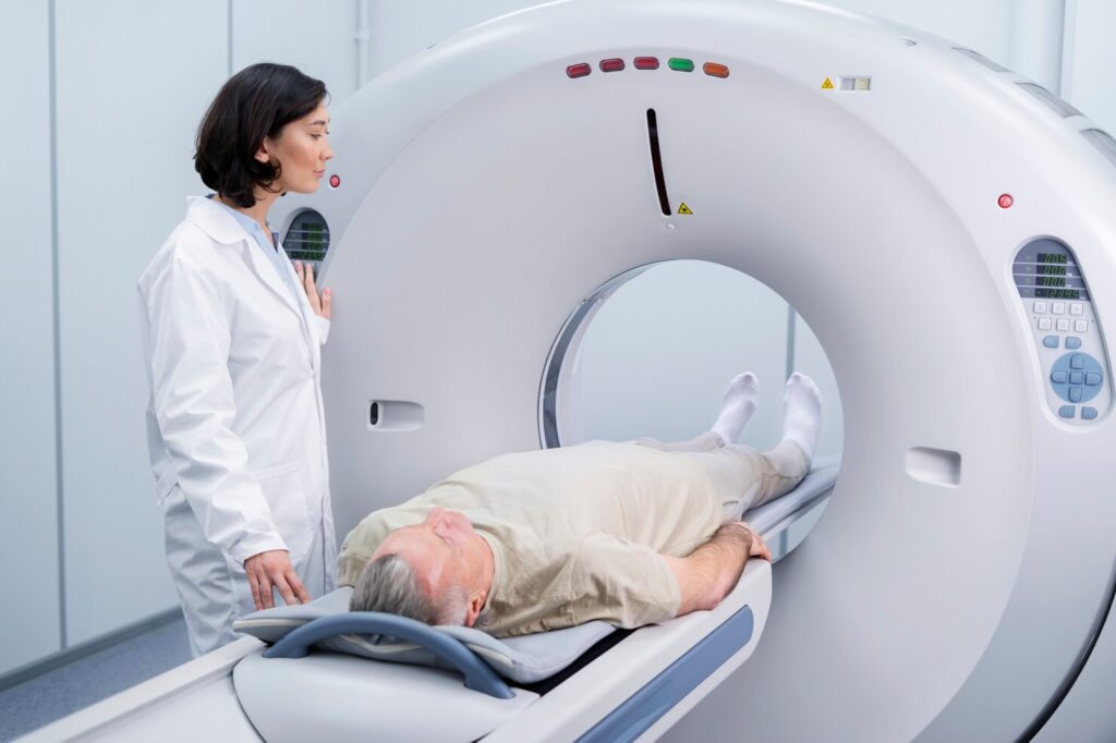
During the scan, patients lie still on a cushioned table that gradually slides into the scanner, with supports used to maintain a comfortable and stable position. The imaging process involves a carefully planned series of sequences, each designed to capture specific information about heart structure, motion, and tissue characteristics. To achieve sharp, diagnostic-quality images, patients are asked to follow a breath-hold protocol. This involves holding the breath for short periods, usually between ten and fifteen seconds, while images are acquired during precise moments of the cardiac cycle.

Breath-holding reduces motion blur caused by chest movement and heartbeat-related displacement, allowing the scanner to capture clearer images of the heart. Clear instructions are delivered through an intercom system, and patients are often given a brief opportunity to practise beforehand. Between breath-holds, normal breathing resumes, providing time to relax and recover. The entire scan typically lasts between forty-five and sixty minutes, depending on the complexity of the assessment and whether contrast-enhanced imaging is required.

Throughout the procedure, patients remain in constant contact with the radiography team, ensuring reassurance and support at all times. A call button is provided so patients can alert staff immediately if assistance is needed. Radiographers monitor progress closely from the control room. Although the scanner produces loud knocking or tapping sounds during image acquisition, these noises are normal and simply indicate that imaging data is being actively collected.

Strategy 3: understanding contrast (gadolinium)

In many cardiac MRI studies, a contrast agent called gadolinium is used to enhance image quality and improve diagnostic detail. Gadolinium is administered through a small intravenous line, usually partway through the scan once initial images have been obtained. After injection, it circulates through the bloodstream and interacts with body tissues predictably, highlighting differences in blood flow, inflammation, and tissue composition that are not visible on non-contrast images. This contrast is particularly valuable for assessing heart muscle viability and detecting scarring or fibrosis within the myocardium. Areas of damaged or diseased tissue absorb gadolinium differently from healthy muscle, allowing cardiologists to identify characteristic patterns that indicate previous injury or ongoing disease processes. 

The amount of contrast used is small, and adverse reactions are rare. Kidney function is often assessed beforehand to ensure the agent can be safely eliminated from the body without risk. Patients typically do not feel any different after receiving gadolinium, aside from a brief cool or mild flushing sensation at the injection site. No sedation or recovery time is usually required following contrast administration. The additional information gained significantly enhances diagnostic accuracy, supports precise clinical decision-making, and helps clinicians develop targeted treatment plans based on reliable, high-quality imaging data.

The result: total diagnostic clarity

The true value of a cardiac MRI lies in the clarity and depth of information it provides. The highly detailed images and quantitative data allow cardiologists to make well-informed decisions about diagnosis, disease severity, and prognosis. Conditions such as cardiomyopathies, inflammatory heart diseases, congenital abnormalities, and ischaemic damage can be assessed comprehensively in a single examination, reducing the need for multiple tests and improving overall diagnostic efficiency.

Based on MRI findings, clinicians can determine whether a patient would benefit from medical management, targeted lifestyle modification, or further intervention. In some cases, the scan may confidently rule out serious disease, providing reassurance and avoiding unnecessary procedures or treatments. In others, it may guide the timing and selection of therapy, including medication adjustments, monitoring strategies, or planning for interventional and surgical approaches. This comprehensive insight supports informed, shared decision-making and proactive heart care. To gain the clearest understanding of your heart health, schedule your cardiac MRI and take a proactive step towards informed, confident decisions about your care.

The silent heart condition: understanding aortic stenosis

Many patients in the early stages of aortic stenosis misinterpret their symptoms as a normal part of ageing. As a result, breathlessness, fatigue, reduced stamina, or slower recovery after activity are often overlooked, delaying recognition of a progressive cardiac condition that requires medical attention. This rationalisation is reinforced by gradual symptom onset and the absence of early pain, often leading individuals to adapt their lifestyles rather than seek assessment. As a result, medical review is frequently delayed, allowing the disease to advance silently until symptoms become more disruptive, persistent, and clinically significant. By the time a diagnosis is made, intervention is often more urgent and complex, carrying higher health risks, fewer treatment options, and less flexibility in selecting the most appropriate therapeutic approach.

The biology: the narrowing of the aortic gateway

The aortic valve acts as the heart’s primary gateway, allowing oxygenated blood to pass from the left ventricle into the aorta and onwards to the rest of the body. In a healthy heart, this valve consists of three thin, flexible leaflets that open fully with each heartbeat and close securely to prevent backflow. With aortic stenosis, these leaflets gradually become thickened, stiffened, and calcified, losing their ability to open freely and efficiently during systole, thereby disrupting normal blood flow dynamics.

Age-related calcium deposition is the most common cause, although congenital valve abnormalities and previous inflammatory conditions can also contribute. As calcification progresses, the effective valve opening becomes smaller, increasing resistance to blood flow leaving the heart. This narrowing forces the left ventricle to work harder to maintain adequate circulation, placing sustained strain on the cardiac muscle over time and accelerating structural changes that compromise performance.

As the valve narrows further, the left ventricle must generate significantly higher pressure to eject blood through the restricted opening. This chronic pressure overload causes the muscular wall of the ventricle to thicken, a process known as left ventricular hypertrophy. While initially compensatory, this thickening reduces ventricular compliance, increases oxygen demand, and eventually compromises filling and pumping efficiency, setting the stage for symptomatic disease, heart failure, and poorer long-term prognosis without timely intervention.

Strategy 1: identifying the “SAD” triad of symptoms

Clinical recognition of aortic stenosis often relies on awareness of the classic symptom cluster known as the “SAD” triad (syncope, angina, and dyspnoea). These symptoms typically emerge as the condition progresses from moderate to severe, indicating that the obstruction has reached a level where the heart can no longer compensate effectively during physical or physiological stress, particularly during exertion or periods of increased demand.

Syncope refers to episodes of fainting or near-fainting, most commonly occurring during exertion or sudden changes in posture. Physical activity increases the body’s demand for blood flow, yet the narrowed valve restricts the heart’s ability to respond appropriately. This mismatch can result in a temporary reduction in blood supply to the brain, causing dizziness, light-headedness, or loss of consciousness, which should always prompt urgent clinical evaluation and specialist assessment.

Angina, or chest pain, occurs when the thickened heart muscle requires more oxygen than the coronary circulation can supply. Even in patients without coronary artery disease, the increased workload and reduced perfusion reserve of the hypertrophied ventricle can lead to chest discomfort. This pain is often exertional, pressure-like in nature, and may resolve with rest as myocardial demand decreases, potentially masking its cardiac origin.

Dyspnoea, defined as shortness of breath on exertion, reflects rising pressures within the heart and lungs. Impaired ventricular filling leads to increased pressure in the left atrium and pulmonary circulation, causing fluid congestion. Over time, activities such as walking uphill, climbing stairs, or carrying groceries become increasingly difficult, reducing independence, exercise tolerance, and overall functional capacity in daily life.

Strategy 2: the Significance of the heart murmur

A heart murmur is frequently the earliest objective sign of aortic stenosis and is often detected during routine clinical examination. General practitioners play a pivotal role in early identification, as careful auscultation can prompt timely referral for further investigation before symptoms become advanced or complications such as heart failure, arrhythmias, or sudden deterioration develop. The characteristic murmur associated with aortic stenosis is described as an ejection systolic murmur. It is generated by turbulent blood flow as blood is forced through the narrowed valve during ventricular contraction. Typically heard best at the upper right sternal edge, the sound may radiate to the carotid arteries in the neck, reflecting altered haemodynamics within the aorta and reduced valve opening.

While the loudness of the murmur does not always correlate with disease severity, its presence is clinically significant, particularly when accompanied by exertional symptoms or risk factors such as advanced age. The murmur serves as an audible marker of altered valve function and highlights the importance of regular cardiovascular examinations as part of routine preventive healthcare and ongoing patient monitoring.

Strategy 3: moving from diagnosis to treatment

Once aortic stenosis is suspected, echocardiography is the definitive diagnostic investigation. This non-invasive ultrasound assessment provides detailed visualisation of valve anatomy, leaflet mobility, and blood flow patterns. By measuring pressure gradients across the valve and calculating the valve area, clinicians can accurately classify disease severity and determine the appropriate timing of intervention, ensuring treatment is neither delayed nor premature. 

Echocardiography also evaluates left ventricular size, wall thickness, and systolic function, offering insight into the heart’s adaptive response to chronic pressure overload. These findings help guide clinical decision-making, particularly in determining whether symptoms are attributable to valve disease and whether intervention is likely to improve survival, functional capacity, and long-term quality of life.

Historically, surgical aortic valve replacement through open-chest surgery was the standard treatment for severe symptomatic disease. Although effective, it carries a higher procedural risk and prolonged recovery for some patients. The advent of transcatheter aortic valve implantation, or TAVI, has revolutionised management by allowing valve replacement via a catheter-based approach, avoiding sternotomy and enabling faster recovery with fewer complications and reduced hospital stays.

Quality of life post-treatment

Following successful valve replacement, most patients experience a significant and sustained improvement in quality of life. Relief from breathlessness and chest discomfort often occurs within weeks, allowing individuals to resume activities that had previously been limited or avoided due to symptoms, fear, or physical exhaustion and declining confidence, while regaining a greater sense of control over their daily routines and physical capabilities. Physical activities such as walking, gardening, and light exercise typically become easier as cardiac output improves and pulmonary congestion resolves. Many patients also report better functional outcomes, sleep quality, improved mood, and increased confidence in their physical abilities, contributing to overall well-being, independence, and renewed social engagement.

Shortness of breath should not be dismissed as a normal part of getting older. It can be an early warning sign of aortic valve disease, where delayed diagnosis may lead to serious complications. Ongoing follow-up is essential to monitor valve function, manage comorbid conditions, and optimise long-term outcomes. Cardiac rehabilitation and clear lifestyle guidance play an important role in supporting recovery and maintaining functional capacity. Timely treatment of aortic stenosis not only improves symptoms but also significantly enhances survival, highlighting the need for early assessment, accurate diagnosis, and patient-centred intervention supported by continuous clinical oversight. Book a valve assessment today to identify potential valve disease early, before symptoms progress.

Heart palpitations vs anxiety: when should you be concerned?

A sudden, forceful thump in the chest that breaks the silence of the night can be deeply unsettling, often prompting an immediate fear that something is seriously wrong with the heart. Heart palpitations are one of the most common reasons people seek urgent medical reassurance, yet they sit within a complex overlap between normal cardiac physiology, heightened bodily awareness, and anxiety-driven responses. While some palpitations are entirely benign and transient, others may signal an underlying rhythm disturbance that warrants further evaluation. The challenge lies in distinguishing between sensations amplified by stress and those arising from genuine electrical irregularities within the heart. Understanding how and why palpitations occur is therefore critical, not only to reduce unnecessary fear but also to ensure that potentially significant cardiac conditions are identified early and managed appropriately.

The biology: your heart’s electrical conduction system

The heart functions through a structured electrical conduction system that maintains a stable rhythm across varying physical and emotional demands. The sinus node, located in the right atrium, acts as the natural pacemaker by generating regular electrical impulses. These impulses travel across the atria, pass through the atrioventricular node, and continue along the His-Purkinje network to the ventricles, resulting in a coordinated and efficient heartbeat. This system is dynamically regulated by the autonomic nervous system to match the body’s needs. Parasympathetic input slows the heart rate during rest, while sympathetic stimulation increases it during physical activity or emotional stress. These adjustments allow rapid adaptation to changes in posture, oxygen demand, and external stressors without compromising cardiac efficiency.

Palpitations occur when normal electrical timing is altered or becomes more noticeable. Ectopic beats, which are extra electrical impulses, are common and often harmless but can create the sensation of a skipped or forceful beat. Anxiety amplifies these sensations by activating the sympathetic nervous system, increasing adrenaline release and heightening awareness of cardiac activity, making benign rhythm changes feel more pronounced.

Strategy 1: deciphering the sensation

Understanding how a palpitation feels provides useful clues about its origin and potential significance. Duration, onset, frequency, and associated symptoms such as breathlessness, dizziness, or chest discomfort refine interpretation and support appropriate triage decisions clinically. Sensation alone cannot diagnose a rhythm disorder, but it helps to differentiate between common benign patterns and those that require further attention through structured clinical assessment and monitoring.

Ectopic beats (PVCs/PACs)

Premature ventricular contractions and premature atrial contractions are among the most frequent causes of palpitations encountered in clinical practice. They are often described as a skipped beat, a brief pause, or a sudden flopping sensation in the chest. The compensatory pause that follows the extra beat allows the heart to fill more fully, making the next contraction feel forceful. In the absence of structural heart disease, these ectopic beats are usually harmless, despite how dramatic or recurrent they may feel to patients.

Tachycardia

 A racing heart creates a distinctly different experience that is often more sustained and noticeable. Sinus tachycardia refers to a fast but regular rhythm driven by the sinus node, commonly associated with anxiety, physical exertion, dehydration, pain, or infection. Supraventricular tachycardia originates from abnormal electrical circuits above the ventricles and is characterised by sudden onset and termination. Patients may notice pounding in the neck, light-headedness, or a sense of internal vibration, reflecting rapid atrial contraction and altered blood flow perception internally.

Atrial fibrillation

Atrial fibrillation produces an “irregularly irregular” rhythm, with no predictable pattern between beats and variable pulse strength. Chaotic electrical activity in the atria replaces organised contraction, leading to inconsistent ventricular responses and reduced cardiac efficiency. This matters because it can impair blood flow and promote clot formation within the atria, increasing the risk of stroke and heart failure. Risk assessment considers age, symptoms, and comorbidities to guide decisions around anticoagulation and long-term rhythm or rate control. Early identification improves outcomes and significantly reduces preventable complications overall.

Strategy 2: identifying external triggers (zeitgebers)

Palpitations are frequently influenced by external and internal timing cues that shape physiological rhythms. Identifying these triggers can significantly reduce symptom burden and restore a sense of control for patients. Keeping a simple symptom diary linking activities, intake, and emotional state can reveal patterns that are not immediately obvious and support targeted intervention strategies.

The vicious cycle of anxiety and palpitations

Psychological interpretation plays a central role. An initial palpitation may trigger concern, which heightens vigilance towards the heartbeat. Catastrophic thinking intensifies physiological arousal, making further palpitations more likely. Education about benign rhythms can interrupt this cycle by reframing sensations as uncomfortable but not harmful. This cognitive shift reduces hypervigilance and supports more effective use of relaxation and coping strategies in daily life contexts.

Chemical triggers

Several commonly used substances alter cardiac excitability, often in combination with psychological stressors. Caffeine increases catecholamine release and shortens electrical refractory periods, encouraging ectopic activity. Nicotine stimulates the heart and raises blood pressure, while alcohol can destabilise atrial electrical conduction. Certain over-the-counter medications contain decongestants that act as stimulants and can provoke palpitations in sensitive individuals. Reading labels and seeking professional advice helps to minimise avoidable triggers effectively overall safely.

Strategy 3: The red flags checklist

Context matters when assessing risk. Age, existing medical conditions, and medication use all influence how palpitations should be interpreted. Syncope or near-syncope suggests transient reductions in cerebral blood flow and warrants urgent assessment. Even a brief loss of consciousness during palpitations should be treated as clinically significant and promptly investigated medically. Chest pain may indicate myocardial ischaemia, while exertion-linked rhythm changes suggest compromised cardiac reserve. A family history of sudden cardiac death or cardiomyopathy further increases risk and should not be overlooked. These contextual factors guide urgency and referral pathways appropriately in practice.

Strategy 4: The diagnostic toolkit

Diagnosis is a process rather than a single test, combining clinical history, physical examination, and targeted investigations to build a coherent picture of cardiac rhythm behaviour over time. Careful symptom correlation, risk assessment, and contextual interpretation are essential components of this process. This structured approach reduces unnecessary testing while ensuring serious conditions are not missed or underestimated clinically, particularly in patients with intermittent, stress-related, or activity-linked symptoms where timing is critical.

12-lead ECG

 A standard twelve-lead electrocardiogram records the heart’s electrical activity from multiple anatomical angles, providing a comprehensive baseline assessment of rhythm, rate, and conduction. It also allows evaluation of heart rate variability, conduction intervals, axis deviation, and repolarisation patterns that may predispose to arrhythmia under physiological or emotional stress. These findings inform risk stratification, referral urgency, and follow-up planning decisions carefully and objectively within established clinical frameworks.

Holter monitoring                                                  

Intermittent symptoms often require extended monitoring to capture relevant rhythm disturbances as they occur in real life rather than controlled settings. Holter monitors record continuous cardiac rhythm over twenty-four to forty-eight hours, while longer-term event monitors can be worn for several weeks. Advances in wearable technology enable assessment during normal daily activities, sleep, and exertion, improving symptom correlation, diagnostic yield, and overall clinical accuracy significantly.

When lifestyle changes aren’t enough

 Management decisions balance symptom severity, objective findings, and patient preference, ensuring treatment aligns with both clinical need and long-term quality of life goals. Lifestyle modification remains the foundation of care, with regular aerobic exercise improving autonomic balance and structured breathing techniques reducing sympathetic surges. Consistency, rather than intensity, is key to sustained benefit. When symptoms persist or impair daily functioning, beta-blockers may reduce adrenergic sensitivity, while other pharmacological or interventional therapies are considered selectively. In some cases, clear reassurance alone restores confidence and reduces symptom-focused anxiety.

If your heart rhythm feels wrong, uncertainty can be more distressing than the symptom itself, particularly when episodes recur without explanation, pattern, or identifiable triggers. Objective assessment replaces speculation with clarity, reassurance, and direction. If palpitations persist, evolve, or cause concern, do not leave it to guesswork or self-diagnosis. Book a consultation to ensure your heart health is guided by evidence rather than fear, allowing informed decisions and appropriate reassurance or treatment based on your individual risk profile, clinical findings, and symptoms.

Why see a private cardiologist? The benefits of rapid diagnostics

Waiting for cardiac test results exerts a significant psychological burden, often characterised by persistent anxiety, disrupted concentration, and heightened awareness of physical sensations that would otherwise pass unnoticed. This uncertainty can be as distressing as symptoms themselves, particularly when individuals understand that heart disease may progress silently beneath an outward appearance of health. In cardiovascular medicine, delay carries measurable biological consequences, making a passive wait-and-see approach clinically inappropriate in many circumstances. Cardiac conditions frequently evolve before symptoms become obvious, narrowing treatment options and increasing long-term risk profiles. Early assessment provides clarity, restores a sense of personal control, and enables timely, evidence-based clinical decisions. Rapid access to expert evaluation and diagnostics is not merely a matter of convenience but a critical determinant in preserving heart structure, functional capacity, and overall long-term prognosis.

The Biology: time is muscle and valve health

The heart responds dynamically to increased workload caused by obstructed blood flow or abnormal valve function that disrupts normal haemodynamics. When coronary arteries narrow, valves leak, or valves stiffen, the myocardium compensates by working harder to maintain adequate circulation throughout the body. Initially, this adaptive response maintains cardiac output, allowing individuals to remain relatively asymptomatic despite significant underlying disease processes. This compensatory response is known as myocardial remodelling, a complex cellular and structural process. In pressure overload conditions, such as aortic stenosis, the heart muscle thickens to generate greater contractile force. In volume overload states, including mitral valve regurgitation, cardiac chambers enlarge to accommodate excess blood volume. Although effective in the short term, these structural changes progressively reduce mechanical efficiency and metabolic reserve.

As structural remodelling advances, the heart muscle becomes less elastic and increasingly fibrotic, altering both structure and function. Electrical conduction pathways may be disrupted, increasing vulnerability to atrial and ventricular arrhythmias. Diastolic filling deteriorates, leading to symptoms such as exertional breathlessness, reduced exercise tolerance, and fatigue. Importantly, these pathological changes may persist even after definitive corrective treatment. Delayed diagnosis directly affects long-term outcomes and therapeutic success. Early identification of disease allows intervention before irreversible myocardial damage occurs. Preserving cardiac architecture and valve integrity improves procedural outcomes, expands eligibility for minimally invasive treatments, and significantly enhances functional recovery following medical or interventional therapy.

Strategy 1: the one-stop shop and same-day diagnostics

Private cardiology services frequently adopt an integrated diagnostic model designed to minimise delay while maximising clinical accuracy. This approach consolidates consultation, investigation, and interpretation into a single coordinated visit. Patients undergo a comprehensive clinical assessment followed immediately by targeted diagnostic testing based on presenting symptoms and risk profile.

Electrocardiography provides essential information regarding rhythm abnormalities, conduction disturbances, and evidence of previous myocardial injury or strain. Echocardiography then offers real-time visualisation of chamber dimensions, valve mechanics, and ventricular performance under physiological conditions. Conducted sequentially, these investigations answer specific clinical questions efficiently and coherently.

Same-day reporting fundamentally transforms the diagnostic process. Results are reviewed and explained during the same appointment, enabling informed discussion and immediate clinical decision-making. This reduces uncertainty, prevents unnecessary interim anxiety, and avoids prolonged waiting periods between investigations that can otherwise delay treatment initiation.

The reliability of this model depends heavily on equipment quality and operator expertise. Hospital-grade imaging systems deliver superior spatial resolution and measurement accuracy compared with many portable alternatives. High-quality diagnostics reduce interpretive ambiguity, support confident management decisions, and minimise the risk of missed, underestimated, or misclassified pathology.

Strategy 2: Continuity of care (the consultant-led model)

Contemporary healthcare systems often divide assessment, diagnosis, and treatment among multiple professionals and departments. While operationally efficient, this fragmentation can weaken continuity and dilute clinical accountability. Recognised barriers such as siloed specialisation and heavy clinical workloads further complicate the management of complex cases, highlighting the need for more coordinated, patient-centred models of care. 

In contrast, consultant-led private cardiology prioritises a single specialist overseeing the entire care pathway from first consultation onward. This continuity ensures that diagnostic findings are interpreted within the broader clinical narrative rather than in isolation. The consultant develops a detailed understanding of symptom evolution, imaging results, and individual risk factors over time. Management decisions therefore evolve logically and consistently, guided by longitudinal clinical insight rather than episodic reassessment.

Personalised care is particularly valuable in cardiovascular disease, where the timing of intervention can be as important as the intervention itself. A consultant familiar with the patient can judge when observation remains appropriate and when escalation becomes necessary. This nuanced judgement cannot be replicated through disconnected or time-limited encounters. When invasive or minimally invasive treatment is required, continuity becomes paramount. The cardiologist who establishes the diagnosis is often the same expert performing procedures such as transcatheter valve implantation or structural heart intervention, ensuring seamless translation from diagnostic assessment to definitive treatment.

Strategy 3: access to advanced 3T MRI and CT imaging

Advanced cardiac imaging has transformed diagnostic precision, allowing detailed evaluation of coronary anatomy, myocardial structure, and tissue composition. Private cardiology services frequently provide expedited access to these technologies, avoiding delays that may influence treatment decisions, disease progression, and long-term outcomes.

Cardiac computed tomography enables rapid, non-invasive assessment of coronary calcium burden and non-calcified plaque. Calcium scoring quantifies atherosclerotic risk, guiding preventive strategies before symptoms emerge. Coronary CT angiography further identifies stenoses and plaque morphology with high diagnostic accuracy and reproducibility. Beyond diagnosis, CT imaging supports detailed procedural planning. Accurate anatomical mapping is essential for interventions such as transcatheter valve replacement, reducing complication risk and improving procedural success rates. Precision imaging directly enhances patient safety, device selection, and procedural efficiency.

Three-Tesla cardiac magnetic resonance imaging represents the reference standard for myocardial characterisation. It distinguishes active inflammation, diffuse fibrosis, and established scar tissue, guiding prognosis and therapy in cardiomyopathy, myocarditis, and infiltrative disease with unparalleled diagnostic confidence and clinical relevance.

When to seek a private second opinion

Second opinions are particularly valuable in complex cardiac conditions where treatment decisions carry significant long-term implications for prognosis, lifestyle, and future intervention options. Valvular heart disease often presents such challenges, with management dependent on anatomy, symptom burden, ventricular response, disease progression rate, and individual surgical risk. Divergent recommendations can understandably generate confusion, hesitation, and uncertainty regarding the most appropriate next step.

A private second opinion allows comprehensive reassessment using advanced imaging and specialist expertise within a focused clinical setting. Previous investigations can be reviewed critically, diagnoses confirmed or refined, and alternative treatment strategies explored in detail with full contextual understanding. This process ensures that decisions reflect current evidence, guideline evolution, procedural availability, and contemporary best practice standards. Second opinions are also appropriate when symptoms and test results appear discordant, inconclusive, or unexplained over time. Subtle disease progression or early myocardial dysfunction may not be fully appreciated through routine assessment alone, particularly in borderline cases. Advanced evaluation can clarify risk, identify overlooked pathology, reassess severity, and refine optimal timing of intervention.

Seeking additional expert input is not a rejection of prior care, but a rational extension of responsible health management. It reflects informed self-advocacy in conditions where disease progression may be clinically silent yet irreversible without timely intervention. Specialist review provides clarity, enabling confident, timely, and personalised clinical decisions aligned with long-term cardiac health. In heart care, time is critical, and early access to specialist assessment with rapid, same-day diagnostics can make a decisive difference. Book your private consultation today for rapid, same-day diagnostics.

Clinic Locations

Multiple locations, one standard of exceptional heart care.

Cromwell Hosptal

London

+44 207 183 0920

Opening hours:
Monday and Friday, On Demand

London Heart Centre

London

+44 207 183 0920

Opening hours:
Thursday, 1pm to 4:30pm

St. Bartholomew's (Barts)

London

+44 207 183 0920

Opening hours:
On Demand

Wellington Hospital

London

+44 207 183 0920

Opening hours:
Thursday, 9:30am to 12pm

The logo for the British Cardiovascular Society features a heart outline painted with blue and red brushstrokes, next to the organisation's name written in blue text.
Logo of the British Cardiovascular Intervention Society (BCIS) with bold red letters BCIS flanked by red arrows, and the full society name in black letters below.
Royal College of Physicians logo with a shield featuring a hand, arm, and plant design, alongside the text Royal College of Physicians in green.
NICOR logo with bold white text on a black background and a curved red section at the bottom.