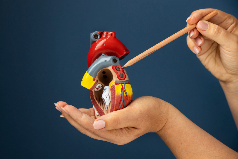
Experiencing a stroke without identifiable risk factors is a uniquely distressing clinical scenario, particularly for younger individuals who would not typically be considered at high cardiovascular risk. When routine investigations fail to reveal a clear cause, the diagnosis of a cryptogenic stroke (stroke without a clear cause) introduces profound uncertainty about recurrence and long-term health. Attention then turns to structural abnormalities within the heart, particularly communications between the upper chambers of the heart (left and right atria) that allow blood clots to bypass the lungs and reach the brain. Among these, patent foramen ovale (PFO) and atrial septal defect (ASD) are most frequently implicated. Although often asymptomatic and easily missed on standard investigations, these conditions can play a decisive role in stroke causation, making precise diagnosis and targeted management essential for effective stroke prevention.
PFO vs. ASD: what is the difference?
A patent foramen ovale (PFO) is a remnant of normal circulation prior to birth, rather than a true structural defect. During foetal life, the foramen ovale allows oxygenated blood from the mother to bypass the inactive lungs. After birth, this “flap” closes to allow normal blood flow through the heart and lungs. However, in around 25% of adults, this closure remains incomplete, leaving a small, flap-like channel that can intermittently open in certain situations, such as during coughing, straining (often called a Valsalva manoeuvre).
An atrial septal defect (ASD) is a true anatomical defect caused by incomplete formation of the wall between the left and right atria. Rather than a flap, there is a persistent opening that allows continuous blood flow between the atria, typically resulting in flow from the left side to the right side, resulting in extra blood flow to the lungs and less flow to the rest of the body. Over time, this abnormal flow can lead to enlargement of the right side of the heart, and, if left untreated, significant problems with the heart and lungs. The distinction between PFO and ASD is clinically important, as each requires a slightly different approach to closure and long-term management.
Accurate differentiation between the two conditions often requires three-dimensional transoesophageal echocardiography (an echo through the gullet) and extends well beyond anatomy, directly informing the most appropriate timing, method, and urgency of intervention for each patient.
The stroke connection: how a clot crosses the heart
The connection between PFO/ASD and stroke is explained by paradoxical embolism, a process in which a blood clot from the legs/pelvis bypasses the lungs and enters the blood flow to the brain and rest of the body. Normally, clots formed in the veins are filtered and broken down in the lungs, preventing them from reaching vital organs.
For patients who have experienced an unexplained stroke, identifying a structural cardiac cause changes the diagnosis from uncertainty to clearly defined and clinically actionable causesThis understanding is crucial, as it allows clinicians to plan appropriate prevention, decide between medical treatment and interventional closure, and offer patients a clear explanation for an event that may previously have seemed entirely without cause.
Diagnosis: beyond the standard ECG
Standard investigations, including ECG, chest X-ray, and basic transthoracic echocardiography (echo), frequently fail to detect smaller defects. A normal initial workup does not exclude a structural cardiac cause, and reliance on first-line tests alone risks overlooking a treatable and directly relevant diagnosis.
A bubble study serves as the primary screening tool in such cases. During echocardiography, microbubbles are injected into a vein and tracked as they enter the right atrium. The appearance of these bubbles in the left atrium within three heart beats indicates the presence of a defect in the heartDiagnostic accuracy is enhanced when the test is performed alongside a Valsalva manoeuvre (essentially straining), which changes pressure in the heart and facilitates the opening of a PFO.
For an accurate diagnosis, a transoesophageal echocardiogram (TOE) remains the investigation of choice. By positioning the ultrasound probe within the oesophagus (gullet), clinicians obtain high-resolution visualisation of the heart structures. This allows for verification of the diagnosis of PFO or ASD, precise measurement of size and confirmation that the defect can be closed by the catheter method. Establishing this distinction is essential before intervention, as closure strategies and device selection vary considerably, and misclassification can directly influence clinical outcomes.
Management and prevention
Management is based on each patient’s clinical profile, including defect size, the presence of symptoms and any history of events such as stroke. For patients with a confirmed PFO and a prior cryptogenic stroke, percutaneous closure is the preferred approach. This minimally invasive, catheter-based procedure places a device, often described as a double-umbrella structure, to seal the opening. PFO closure significantly reduces the risk of recurrent stroke by around 50%The procedure carries a small risk of procedural complications including heart rhythm problems, underscoring the importance of appropriate patient selection and specialist experience.
For ASD, closure is indicated in patients who have had a stroke and also when there is right heart enlargement, with or without symptoms such as breathlessness or fatigue. Depending on anatomy, closure may be achieved percutaneously or via surgical repair. Early intervention prevents progression to right heart failure, and heart rhythm problems .
If you have experienced an unexplained stroke or TIA, or have been told you have a murmur or a hole in your heart, it is important to know that clear answers and effective treatments are available. With the right investigations and specialist care, these conditions can be accurately diagnosed and managed. Schedule an assessment to clarify your diagnosis and explore the most appropriate treatment options.